Chakrabarti 2018 MiP2018
| Fatty acid binding protein 3 regulation of mitochondrial lipids - A strategy for exceptional longevity in the Pipistrelle bat. |
Link: MiP2018
Pollard AK, Ingram TL, Ortori CA, Shephard F, Liddell S, Barrett DA, Chakrabarti L (2018)
Event: MiP2018
It is generally accepted that smaller mammals with higher metabolic rates have shorter maximal lifespans. The very few mammalian species that don’t abide by these rules can give insight into how the detrimental effects of ageing may be delayed. The recorded maximum lifespans of micro-bats are exceptional, reaching over 40 years, compared with the similarly-sized laboratory mouse of 4 years. We investigated the possibility that differences in the biochemical composition of mitochondria might be associated with maximal life-span differences between bat and mouse species. We used 2D gel electrophoresis for proteomics and ultra-high performance liquid chromatography coupled with high resolution mass spectrometry lipidomics, to interrogate mitochondrial fractions prepared from Mus musculus and Pipistrelle pipistrellus brain and skeletal muscle. Identified potential modifiers of mitochondrial ageing were further investigated in Caenorhabditis elegans RNAi knock-downs for lipid binding proteins 4, 5 and 6. We show that mitochondrial proteomes are distinctly different when comparing mouse with bat, and their mitochondrial lipid signatures clearly define the tissue and species of origin. In the bat high levels of free fatty acids and N-acylethanolamine lipid species together with a significantly greater abundance of fatty acid binding protein 3 in muscle (1.8 fold, p=0.037) comprise a pathway which may be important for increased longevity in mammals. Manipulation of fatty acid binding protein orthologues in C.elegans confirmed these proteins connect mitochondrial function and lifespan. Our comparison is the first to delineate mitochondrial profiles in the bat to reveal an intrinsic biochemistry consistent with an enhanced ability to counter detrimental effects of ageing[1–4].
• Bioblast editor: Plangger M, Kandolf G
Labels: MiParea: mt-Membrane, Comparative MiP;environmental MiP Pathology: Aging;senescence
Organism: Mouse, Other mammals, Caenorhabditis elegans Tissue;cell: Skeletal muscle, Nervous system
Regulation: Fatty acid
Affiliations
Pollard AK(1), Ingram TL(1), Ortori CA(3), Shephard F(1), Liddell S(2), Barrett DA(3), Chakrabarti L(1,4)
- School Veterinary Medicine Science
- School Biosciences
- Centre Analytical Bioscience, School Pharmacy; Univ Nottingham, Sutton Bonington, UK
- MRC-ARUK Centre Musculoskeletal Ageing. - [email protected]
Figures
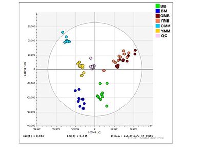
Figure 1. Mitochondrial lipid composition differs between the bat and mouse mitochondrial proteomes. Orthogonal partial least square-discriminant analysis (OPLS-DA) of lipids found in the brain and skeletal muscle mitochondria from the mouse and the bat. Separation across the x-axis is according to tissue type with the skeletal muscle mitochondrial samples congregating to the left quadrants and the brain mitochondrial samples to the right. Along the y-axis separation delineates mammalian species with the bat mitochondrial samples grouping at the lower quadrants and the mouse mitochondrial samples grouping at the upper quadrants. Bat brain (BB) mitochondrial samples (adult, n=10) are shown on the OPLS-DA by the green circles. Bat skeletal muscle (BM) mitochondria (adult, n=10) are indicated by the dark blue circles. Young mouse brain (YMB) mitochondria aged 4-11 weeks (n=10) and aged mouse brain mitochondria (OMB) aged 78 weeks (n=10) are denoted by orange and red circles respectively. Young mouse skeletal (YMM) muscle mitochondria aged 4-11 weeks (n=9) and aged mouse skeletal muscle mitochondria (OMM) aged 78 weeks (n=10) are indicated by the yellow and blue circles, respectively.
References
- Pollard A, Shephard F, Freed J, Liddell S, Chakrabarti L (2016) Mitochondrial proteomic profiling reveals increased carbonic anhydrase II in aging and neurodegeneration. Aging (Albany NY) 8:2425-36.
- Pollard AK, Ortori CA, Stöger R, Barrett DA, Chakrabarti L (2017) Mouse mitochondrial lipid composition is defined by age in brain and muscle. Aging (Albany NY) 9:986-98.
- Ingram T, Chakrabarti L (2016) Proteomic profiling of mitochondria: what does it tell us about the ageing brain? Aging (Albany NY) 8:3161-79.
- Foley NM, Hughes GM, Huang Z, Clarke M, Jebb D, Whelan CV, Petit EJ, Touzalin F, Farcy O, Jones G, Ransome RD, Kacprzyk J, O'Connell MJ, Kerth G, Rebelo H, Rodrigues L, Puechmaille SJ, Teeling EC (2018) Growing old, yet staying young: The role of telomeres in bats’ exceptional longevity. Sci Adv 4:eaao0926.


