Kim 2020 Cells
| Kim J, Cheong JH (2020) Role of mitochondria-cytoskeleton interactions in the regulation of mitochondrial structure and function in cancer stem cells. Cells 9:1691. https://doi.org/10.3390/cells9071691 |
Abstract: Despite the promise of cancer medicine, major challenges currently confronting the treatment of cancer patients include chemoresistance and recurrence. The existence of subpopulations of cancer cells, known as cancer stem cells (CSCs), contributes to the failure of cancer therapies and is associated with poor clinical outcomes. Of note, one of the recently characterized features of CSCs is augmented mitochondrial function. The cytoskeleton network is essential in regulating mitochondrial morphology and rearrangement, which are inextricably linked to its functions, such as oxidative phosphorylation (OXPHOS). The interaction between the cytoskeleton and mitochondria can enable CSCs to adapt to challenging conditions, such as a lack of energy sources, and to maintain their stemness. Cytoskeleton-mediated mitochondrial trafficking and relocating to the high energy requirement region are crucial steps in epithelial-to-mesenchymal transition (EMT). In addition, the cytoskeleton itself interplays with and blocks the voltage-dependent anion channel (VDAC) to directly regulate bioenergetics. In this review, we describe the regulation of cellular bioenergetics in CSCs, focusing on the cytoskeleton-mediated dynamic control of mitochondrial structure and function.
• Bioblast editor: Gnaiger E
Hydrogen ion ambiguities in the electron transfer system
Communicated by Gnaiger E (2023-10-08) last update 2023-11-10
- Electron (e-) transfer linked to hydrogen ion (hydron; H+) transfer is a fundamental concept in the field of bioenergetics, critical for understanding redox-coupled energy transformations.
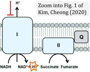
- However, the current literature contains inconsistencies regarding H+ formation on the negative side of bioenergetic membranes, such as the matrix side of the mitochondrial inner membrane, when NADH is oxidized during oxidative phosphorylation (OXPHOS). Ambiguities arise when examining the oxidation of NADH by respiratory Complex I or succinate by Complex II.
- Oxidation of NADH or succinate involves a two-electron transfer of 2{H++e-} to FMN or FAD, respectively. Figures indicating a single electron e- transferred from NADH or succinate lack accuracy.
- The oxidized NAD+ is distinguished from NAD indicating nicotinamide adenine dinucleotide independent of oxidation state.
- NADH + H+ → NAD+ +2{H++e-} is the oxidation half-reaction in this H+-linked electron transfer represented as 2{H++e-} (Gnaiger 2023). Putative H+ formation shown as NADH → NAD+ + H+ conflicts with chemiosmotic coupling stoichiometries between H+ translocation across the coupling membrane and electron transfer to oxygen. Ensuring clarity in this complex field is imperative to tackle the apparent ambiguity crisis and prevent confusion, particularly in light of the increasing number of interdisciplinary publications on bioenergetics concerning diagnostic and clinical applications of OXPHOS analysis.
Labels: Pathology: Cancer


