Difference between revisions of "Kimoloi 2019 MiPschool Coimbra"
(Created page with "{{Abstract}} {{Labeling}}") |
|||
| Line 1: | Line 1: | ||
{{Abstract}} | {{Abstract | ||
|title=[[Image:MiPsocietyLOGO.JPG|left|90px|Mitochondrial Physiology Society|MiPsociety]] | |||
|info=[[MitoEAGLE]] | |||
|authors= | |||
|year=2019 | |||
|event=MiPschool Coimbra 2019 | |||
|abstract=[[Image:MITOEAGLE-logo.jpg|left|100px|link=http://www.mitoeagle.org/index.php/MitoEAGLE|COST Action MitoEAGLE]] | |||
|editor=[[Plangger M]], | |||
}} | |||
{{Labeling}} | {{Labeling}} | ||
== Affiliations == | |||
Kimoloi Sammy1, Baris OR 1, Wiesner RJ.1,3,4 | |||
1Center for Physiology and Pathophysiology, Institute of Vegetative Physiology, Medical Faculty, University of Köln, 50931 Köln, Germany | |||
3 Center for Molecular Medicine Cologne, CMMC, University of Köln, 50931 Köln, Germany | |||
4 Cologne Excellence Cluster on Cellular Stress Responses in Ageing-associated Diseases (CECAD), 50674 Köln, Germany | |||
== Figures == | |||
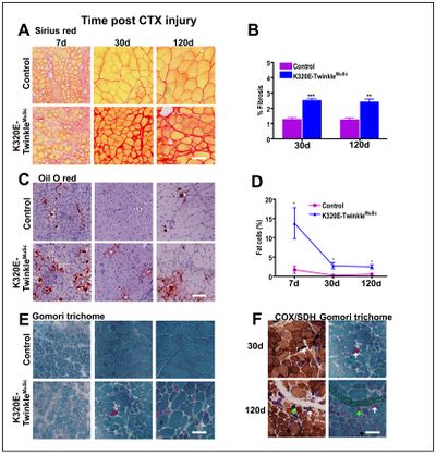
[[File:Kimoloi_Figure1.jpg|left|400 px]] Figure 1: Mitochondrial DNA maintainance defecfects in MuSc results in fibrosis, fat infiltration and ragged red phenotype in regenerated muscles. A and B) Sirius red staining analysis showing increased fibrosis at 30 and 120d post CTX injury, C and D) oil O red showing increased adipocyte infiltration in mutant mice at 30 and 120d post CTX injury, E) modified Gomori trichrome staining indicating presence of RRFs in mutant mice, F) serial sections stained for COX-SDH and Gomori trichrome. **p<0.01 t-test *p<0.05 two-way ANOVA, Scale bar=100µm. | |||
== References == | |||
Mauro, A., Satellite cell of skeletal muscle fibers. J Biophys Biochem Cytol, 1961. 9: p. 493-5. | |||
2. Brack, A.S. and P. Munoz-Canoves, The ins and outs of muscle stem cell aging. Skelet Muscle, 2016. 6: p. 1. | |||
3. Sousa-Victor, P., et al., Muscle stem cell aging: regulation and rejuvenation. Trends Endocrinol Metab, 2015. 26(6): p. 287-96. | |||
4. Baris, O.R., et al., Mosaic Deficiency in Mitochondrial Oxidative Metabolism Promotes Cardiac Arrhythmia during Aging. Cell Metab, 2015. 21(5): p. 667-77. | |||
Revision as of 15:22, 27 June 2019
Link: MitoEAGLE
(2019)
Event: MiPschool Coimbra 2019
• Bioblast editor: Plangger M
Labels:
Affiliations
Kimoloi Sammy1, Baris OR 1, Wiesner RJ.1,3,4 1Center for Physiology and Pathophysiology, Institute of Vegetative Physiology, Medical Faculty, University of Köln, 50931 Köln, Germany 3 Center for Molecular Medicine Cologne, CMMC, University of Köln, 50931 Köln, Germany 4 Cologne Excellence Cluster on Cellular Stress Responses in Ageing-associated Diseases (CECAD), 50674 Köln, Germany
Figures
Figure 1: Mitochondrial DNA maintainance defecfects in MuSc results in fibrosis, fat infiltration and ragged red phenotype in regenerated muscles. A and B) Sirius red staining analysis showing increased fibrosis at 30 and 120d post CTX injury, C and D) oil O red showing increased adipocyte infiltration in mutant mice at 30 and 120d post CTX injury, E) modified Gomori trichrome staining indicating presence of RRFs in mutant mice, F) serial sections stained for COX-SDH and Gomori trichrome. **p<0.01 t-test *p<0.05 two-way ANOVA, Scale bar=100µm.
References
Mauro, A., Satellite cell of skeletal muscle fibers. J Biophys Biochem Cytol, 1961. 9: p. 493-5. 2. Brack, A.S. and P. Munoz-Canoves, The ins and outs of muscle stem cell aging. Skelet Muscle, 2016. 6: p. 1. 3. Sousa-Victor, P., et al., Muscle stem cell aging: regulation and rejuvenation. Trends Endocrinol Metab, 2015. 26(6): p. 287-96. 4. Baris, O.R., et al., Mosaic Deficiency in Mitochondrial Oxidative Metabolism Promotes Cardiac Arrhythmia during Aging. Cell Metab, 2015. 21(5): p. 667-77.


